
Fig. 7
- ID
- ZDB-FIG-180413-28
- Publication
- Dunn et al., 2017 - Dual Roles of Fer Kinase Are Required for Proper Hematopoiesis and Vascular Endothelium Organization during Zebrafish Development
- Other Figures
- All Figure Page
- Back to All Figure Page
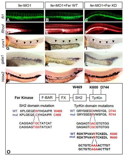
Kinase-inactive Fer rescues vascular tubulogenesis but not hematopoiesis gene expression defects. The injection of fer mRNA and fer-MO1 into fli1a::nEGFP embryos results in increased cell migration into the ISV regions ((B)—WT, (C)—kinase inactive (KD)) when compared to fer-MO1 alone (A), and rescues tube formation ((E)—WT, (F)—KD, Rhodamine-Dextran circulation after 15 min, when compared to fer-MO1 alone (D)). Wild type Fer ameliorates defects in erythrocyte expansion, as observed with runx1 (H), gata1 (K) and hbbe2 (N), while inactive Fer does not ((I,L,P) respectively). fer-MO1 alone is shown in (G,J,M) respectively. Embryos were examined at 30 somites (~28 hpf). Domains of Fer kinase in zebrafish, showing the highly conserved tryptophan and aspartic acid residues, in the SH2 and tyrosine kinase domains, respectively, are shown. Mutating either tryptophan to cysteine (W469C), aspartic acid to arginine (D744R) or lysine to arginine (K600R) resulted in the loss of kinase activity for Fer. The amino acid sequence is shown on top, with the corresponding DNA sequence changes below ((O), Fer KD expression confirmation shown in Supplementary Figure S1). |