
Fig. 5
- ID
- ZDB-FIG-180413-26
- Publication
- Dunn et al., 2017 - Dual Roles of Fer Kinase Are Required for Proper Hematopoiesis and Vascular Endothelium Organization during Zebrafish Development
- Other Figures
- All Figure Page
- Back to All Figure Page
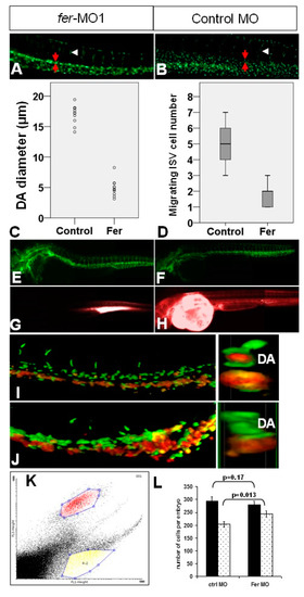
The loss of Fer kinase function resulted in vascular tubulogenesis defects. The loss of Fer kinase function decreased the diameter of the dorsal aorta (A,C) and resulted in fewer migrating cells into the ISV regions (D) at 30 somites (Control embryos (B–D). Rhodamine–Dextran injections into DA resulted in no visible dye circulation in Fer deficient embryos ((E), fli1::nEGFP, (G), Rhodamine), when compared to control embryos ((F), fli1::nEGFP, (H), Rhodamine). Confocal microscopy images demonstrated a concurrent loss of vasculature organization in the region of the DA and gain of hematopoietic precursors at 26 hpf ((I), control MO, (J), fer-MO1). The right images in (I,J) are digital cross-sections, taken approximately midway through the image; z-stacks showed no gata1::dsRed signal in the region that should be the inner-DA. The FACS analysis showed a shift in the number of gata1::dsRed cells, along with a loss of vascular endothelial population ((K), gated regions of non-overlapping, fli1a::nEGFP and gata1::dsRed cells, (L), quantitative plot showing differences in the cell population from (K) in the presence/absence of Fer kinase activity). |
| Fish: | |
|---|---|
| Knockdown Reagent: | |
| Observed In: | |
| Stage: | Prim-5 |