
Fig. 4
- ID
- ZDB-FIG-180412-17
- Publication
- Ribeiro et al., 2017 - Foxj1a is expressed in ependymal precursors, controls central canal position and is activated in new ependymal cells during regeneration in zebrafish
- Other Figures
- All Figure Page
- Back to All Figure Page
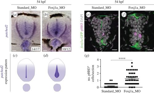
Shh signalling and proliferation are enhanced in the absence of Foxj1a. (a,b) Expression of patched2 by ISH in 54 hpf embryos injected with Standard Morpholino (MO) (a) or foxj1a MO (b). Two types of expression patterns were observed, which are schematically represented in (c,d). The majority of foxj1a morphants display increased patched2 expression in ventral cells surrounding the lumen. (e,f) Confocal images of transverse sections of Tg(0.6foxj1a:GFP) 54 hpf embryos injected with Standard MO (e) or foxj1a MO (f) and immunostained against phospho-histone H3 (pHH3) to detect mitotic cells (magenta). The foxj1a:GFP reporter is shown in green and DAPI-labelled nuclei are shown in grey. Scale bars, 10 µm. (g) Quantification of the number of pHH3+ cells per section (Standard MO, n = 43; foxj1a MO, n = 46). Data obtained from three independent experiments. Each point represents the average of two non-consecutive sections per embryo and the mean and s.d. bars are also shown. p value calculated using the two-tailed unpaired t-test (****p < 0.0001). |