
Fig. 3
- ID
- ZDB-FIG-180412-16
- Publication
- Ribeiro et al., 2017 - Foxj1a is expressed in ependymal precursors, controls central canal position and is activated in new ependymal cells during regeneration in zebrafish
- Other Figures
- All Figure Page
- Back to All Figure Page
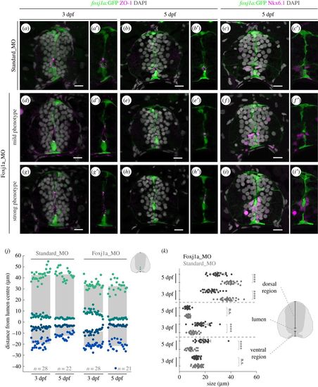
Neural tube lumen closure is perturbed in the absence of Foxj1a. (a–i′) Representative images of transverse sections of the neural tube of Tg(0.6foxj1a:GFP) transgenic larvae injected with Standard Morpholino (MO) (a–c′) or foxj1a MO (d–i′) at one-cell stage and analysed at 3 and 5 dpf. foxj1a MO-injected larvae showed lumen closure phenotypes with variable degrees of severity, between mild (d–f′) and strong (g–i′), as highlighted by the apical ZO-1 immunostaining (magenta) (a,b′,d,e′,g,h′). (c,c′,f,f′,i,i′) Nkx6.1+ cells (magenta) are still present lining the ventral half of the central canal in foxj1a morphants. DAPI-labelled nuclei are shown in grey. Scale bars: 10 µm. (j) Quantification of lumen closure in Standard MO and foxj1a MO larvae at 3 and 5 dpf. The positions are normalized to the middle point of the lumen. Sample number is shown in the graph and includes data from three independent experiments. (k) Quantification of the size of the lumen and the ventral and dorsal regions, derived from the data shown in (j). Each point represents one individual and the mean and s.d. bars are also shown. p values calculated using two-tailed unpaired t-test (****p < 0.0001; n.s., not significant). |