
Fig. s1
- ID
- ZDB-FIG-180410-9
- Publication
- Nagashima et al., 2017 - Anisotropic Müller glial scaffolding supports a multiplex lattice mosaic of photoreceptors in zebrafish retina
- Other Figures
- All Figure Page
- Back to All Figure Page
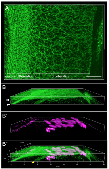
Maximum intensity z-projection and lateral slice view of retinal margin illustrating shape and position of mitotic figures. (A) Snap shot from a 3D reconstruction of the retinal margin and retinal pigmented epithelium (RPE) in a flat-mount preparation immunostained for ZO1 (green). The front of the eye is to the right. Proliferative, differentiating (pre-column), and differentiated (mature mosaic) zones of the retina are labeled. (B-B”) Lateral view (xz plane) of ZO1 (green) and the mitotic marker, pH3 (magenta). (B) The RPE (arrow) and neural retinal epithelium (arrowhead) are separate but closely apposed, especially in the proliferative zone. (B’) The pH3+ mitotic cells are tilted with respect to the surface of the retinal epithelium, and the resultant parallax precludes identifying the profiles of pH3+ mitotic cells in the ZO1 channel in a maximum intensity z-projection (e.g., Fig. 1G'). (B’’) Proliferating endothelial cells in the circumferential blood vessel (yellow arrow) of the vitreous circulation, which lies below the retinal germinal zone [1], are also pH3+ (asterisks). Scale bar: 20 μm (A). |