
FIGURE
Fig. s5
- ID
- ZDB-FIG-180410-12
- Publication
- Nagashima et al., 2017 - Anisotropic Müller glial scaffolding supports a multiplex lattice mosaic of photoreceptors in zebrafish retina
- Other Figures
- All Figure Page
- Back to All Figure Page
Fig. s5
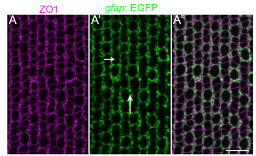
Müller glial apical processes are preferentially distributed into parallel, inter-column bands. (A-A”) Retinal flat-mount immunocytochemistry for ZO1 (magenta) in the Müller glial transgenic reporter line, Tg(gfap: EGFP). Müller glial processes (GFP+) completely surround the profiles of individual rods and cones, forming relatively thin intracolumn lamellae (short arrow), and thicker inter-column expansions (long arrow). Refer to Fig 1 A and B for a description of columns. Scale bar: 10 μm. |
Expression Data
Expression Detail
Antibody Labeling
Phenotype Data
Phenotype Detail
Acknowledgments
This image is the copyrighted work of the attributed author or publisher, and
ZFIN has permission only to display this image to its users.
Additional permissions should be obtained from the applicable author or publisher of the image.
Full text @ Neural Dev.