
FIGURE
Fig. 1
- ID
- ZDB-FIG-180405-3
- Publication
- Matsubara et al., 2017 - New photic stimulating system with white light-emitting diodes to elicit electroretinograms from zebrafish larvae
- Other Figures
- All Figure Page
- Back to All Figure Page
Fig. 1
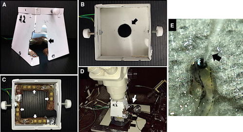
A built-in light-emitting diode (LED) light stimulator. a External view and b internal view from the upper part of the LED light stimulator. The partition plate has a circular hole of 2.0 cm in diameter (arrow). c The inside of the partition plate is removed. Twelve white LEDs are installed in the lower part of the stimulator (arrows). All LEDs are covered with copper netting. d The stimulator can be attached to the lower part of a stereomicroscope. e The view of larvae and a glass microelectrode (arrow) with a stereomicroscope. The damp paper towel which covered the larva’s body is removed |
Expression Data
Expression Detail
Antibody Labeling
Phenotype Data
Phenotype Detail
Acknowledgments
This image is the copyrighted work of the attributed author or publisher, and
ZFIN has permission only to display this image to its users.
Additional permissions should be obtained from the applicable author or publisher of the image.
Full text @ Doc. Ophthalmol.