
Fig. S5
- ID
- ZDB-FIG-180222-28
- Publication
- Akerberg et al., 2017 - Histone demethylases Kdm6ba and Kdm6bb redundantly promote cardiomyocyte proliferation during zebrafish heart ventricle maturation
- Other Figures
- All Figure Page
- Back to All Figure Page
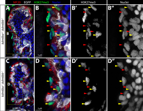
kdm6ba/bb are not required for the transient depletion of bulk H3K27me3 levels in trabeculating cardiomyocytes. (A-D”) Confocal microscopy immunofluorescence images of sections through the hearts of 3 dpf Tg(kdrl:EGFP) control and kdm6ba/bb-deficient embryos stained with antimyosin heavy chain (red, MF20, myocardium), anti-EGFP (white, endocardium), and anti-H3K27me3 (green) antibodies with Hoechst-stained nuclei in blue (A, B, C, D). Yellow boxed areas in A and C are shown zoomed in B-B” and D-D” respectively with grey-scale single channel images of H3K27me3 staining (B’, D’) and nuclei (B”, D”). Arrowheads indicate myocardial cells with robust (yellow) or depleted (red) bulk H3K27me3 levels. 20 μM and 5 μM scale bars are shown |
Reprinted from Developmental Biology, 426(1), Akerberg, A.A., Henner, A., Stewart, S., Stankunas, K., Histone demethylases Kdm6ba and Kdm6bb redundantly promote cardiomyocyte proliferation during zebrafish heart ventricle maturation, 84-96, Copyright (2017) with permission from Elsevier. Full text @ Dev. Biol.