
Fig. 5
- ID
- ZDB-FIG-180208-21
- Publication
- Dudczig et al., 2017 - Developmental and adult characterization of secretagogin expressing amacrine cells in zebrafish retina
- Other Figures
- All Figure Page
- Back to All Figure Page
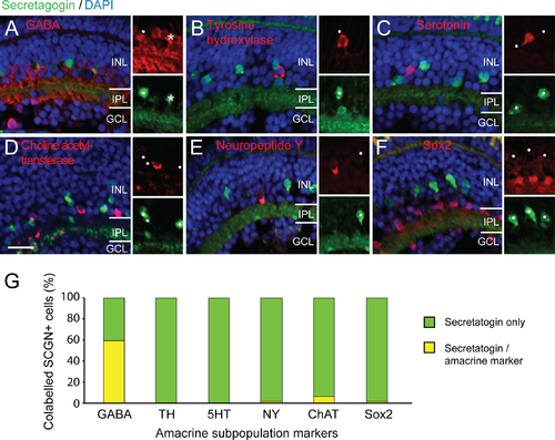
Secretagogin amacrine subtype markers and other calcium binding proteins within the zebrafish retina. (A–F) Micrographs showing cross-sections through zebrafish retina at 5 days postfertilization. Insets show separate red and green channels of subregion of the double channel images. Co-labeling (asterisks) of secretagogin—SCGN (green) with amacrine subtypes markers (red): GABA (A), tyrosine hydroxylase–TH (B), serotonin– 5HT (C), choline acetyltransferase–ChAT (D), neuropeptide Y—NY (E), Sox2 (F). Most SCGN+ cells show little co-localization with other markers (white dots). (G) Graph shows proportion of secretagogin cells that also co-label for the other amacrine markers (asterisks). Scale bar (D) for A–F is 20 μm. |