
FIGURE
Fig. 2
- ID
- ZDB-FIG-180208-18
- Publication
- Dudczig et al., 2017 - Developmental and adult characterization of secretagogin expressing amacrine cells in zebrafish retina
- Other Figures
- All Figure Page
- Back to All Figure Page
Fig. 2
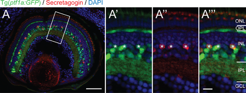
Secretagogin positive cells co-label with the Ptf1a:GFP amacrine marker in the inner nuclear layer. Micrograph at 5 days postfertilization showing secretagogin immunostained Tg(ptf1a:GFP) zebrafish retinas. Higher magnification of boxed view of boxed inset shows co-localization (asterisks) of SCGN+ (red) and Ptf1a:GFP+ (green) within individual cells marked in the green (A'), red (A'') and double (A''') channels.). ONL: outer nuclear layer; OPL: outer plexiform layer; INL: inner nuclear layer; IPL: inner plexiform layer; GCL: ganglion cell layer. Scale bar (A) is 50 μm, scale bar (A''') for A'–A''' is 10 μm. |
Expression Data
Expression Detail
Antibody Labeling
Phenotype Data
Phenotype Detail
Acknowledgments
This image is the copyrighted work of the attributed author or publisher, and
ZFIN has permission only to display this image to its users.
Additional permissions should be obtained from the applicable author or publisher of the image.
Full text @ PLoS One