
Fig. 5
- ID
- ZDB-FIG-180206-26
- Publication
- Bower et al., 2017 - SERCA directs cell migration and branching across species and germ layers
- Other Figures
- All Figure Page
- Back to All Figure Page
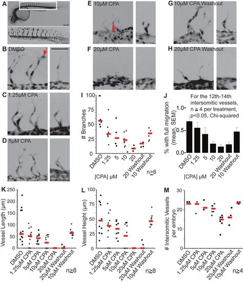
SERCA function controls the rates of mesodermal migration and budding. (A) Wild-type 28 hpf Tg(kdrl:eGFP) zebrafish embryo demonstrates intersomitic vasculature seen by wide field (top, scale bar: 200 μm) and higher magnification below (scale bar: 100 μm). For A-H, images are inverted for improved clarity. (B-H) Paired confocal images show 3D reconstructions of the 14th-15th and 20th intersomitic vessels from embryos treated with (B) DMSO, (C) 1.25 µM CPA, (D) 5 µM CPA, (E) 10 µM CPA, (F) 20 µM CPA (G) 10 µM CPA washed out after 2 h, and (H) 20 µM CPA washed out after 2 h. Varying CPA dose results in dose-dependent reduction in vascular budding until at 20 μM, budding is reversibly suspended. Budding resumes after CPA washout. Scale bar: 50 µm for all images. Red arrows indicate distal-most positioning of nuclei in B and E (see caption for I,J). (I,J) Plots show, for each treatment, the collective number of branches (red lines are medians) on vessels 13-16 which form in the middle of the treatment (I) and the proportion of branches with cell nuclei at tip positions (J): compare the positions of nuclei (arrowed) in B versus E. Error bars in J indicate the s.e.m. In a parallel dose-dependent manner, CPA reduces branch numbers (I) and distal migration of endothelial cells (J) with resumption of branching and migration upon CPA washout. (K-M) Vessel path length (K), linear vessel height (L), and the total number of intersomitic vessels per embryo (M) measured in 3D show similar CPA dose- and time-dependent reductions in branching. Data in K and L are shown for the 20th intersomitic vessel, which forms during the treatment (red lines are medians). |