
Fig. 5
- ID
- ZDB-FIG-180105-26
- Publication
- Wu et al., 2017 - Fine-tune regulation of carboxypeptidase N1 controls vascular patterning during zebrafish development
- Other Figures
- All Figure Page
- Back to All Figure Page
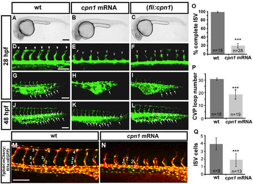
Overexpression of cpn1 causes vascular defects in zebrafish embryos. (A–C) The bright field images of uninjected control and cpn1 mRNA-injected (0.93ng) and (fli1:cpn1) embryos at 28 hpf. Compared with controls (arrowheads in (D), arrows in (G), the overexpression of cpn1 caused ISV growth defects (hollow arrowheads in (E, F) and less honeycomb-like structure formation in CVP at 28 hpf (H,I). At 28 hpf, ISVs reached DLAV and formed a honeycomb-like structure in CVP in controls. At 48 hpf, CVP loop formation was less (arrows in K, L) than in controls (arrows in J). (M,N) Injection of cpn1 mRNA into Tg(kdrl:mCherry ci5; fli1a:nEGFP y7) embryos (N) shows that the cell number per ISV was lower than in control (M) and ISVs did not reach DLAV as compared to control at 28hpf. (O) The percentage of completed ISVs decreased by approximately 80% in cpn1 mRNA injected embryos (n = 18 in wt and n = 28 in cpn1 mRNA) at 28 hpf. (P) The loop formation at CVP decreased in cpn1 mRNA injected samples (n = 18 in wt and n = 19 in cpn1 mRNA) at 48 hpf. (Q) The average number of cells per ISV decreased in cpn1 mRNA injected embryos (n = 8 in wt and n = 15 in cpn1 mRNA) at 30 hpf. The images are the representative pictures from two independent experiments. Quantitative data are represented as means ± S.D. ***Refers to p < 0.0001 by an unpaired Student’s t-test. Scale bars are 200 µm for A–C and 100 µm for D–N. |