
Fig. 8
- ID
- ZDB-FIG-171228-45
- Publication
- Biechl et al., 2017 - Identification of accessory olfactory system and medial amygdala in the zebrafish
- Other Figures
- All Figure Page
- Back to All Figure Page
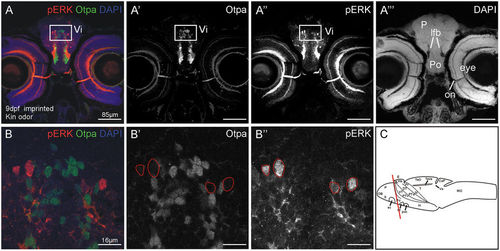
Example of pERK activation and Otpa-positive cells in the intermediate nucleus of the ventral telencephalon (Vi) and indication of the sector that was counted. (A–A'') Confocal photomicrographs show in addition to pERK, the nuclear stain DAPI and Otpa. (B) Higher power details of insert show pERK and Otpa-positive cells in confocal photography in histological material of tested fish. (C) Larval brain in lateral view shows section level. Abbreviations: ac: anterior commissure; CeP: cerebellar plate; DT: dorsal thalamus (thalamus); E: epiphysis; EmT: eminentia thalami; GL: glomerular layer; H: hypothalamus; Ha: habenula; INL: inner nuclear layer; lfb: lateral forebrain bundle; mdG2: mediodorsal glomerulus 2; MO: medulla oblongata; N: nucleus of the medial longitudinal fascicle; OB: olfactory bulb; on: optic nerve; P: pallium; Po: preoptic region; Pr: pretectum; PTd, PTv: dorsal, ventral part of posterior tuberculum; S: subpallium; T: tegmentum; TeO: optic tectum; TeVe: tectal ventricle; Va: valvula cerebelli; VT: ventral thalamus (prethalamus). |