
Fig. 6
- ID
- ZDB-FIG-171228-44
- Publication
- Biechl et al., 2017 - Identification of accessory olfactory system and medial amygdala in the zebrafish
- Other Figures
- All Figure Page
- Back to All Figure Page
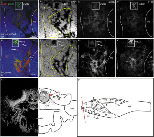
xample of pERK activation in olfactory bulb section containing mdG2 (frame) in imprinted and non-imprinted zebrafish larva. (A–A''') confocal photomicrograph of a sectioned imprinted larva. (B–B''') confocal photomicrograph of a sectioned non-imprinted larva. Channels comprise in addition to pERK, the nuclear stain DAPI and the calcium-binding protein immunostain S100. (C) Shows DAPI (left) and a schema with olfactory bulb fields that were counted (mdG2, INL, GL). (D) Larval brain in lateral view shows section level. Abbreviations: ac: anterior commissure; CeP: cerebellar plate; DT: dorsal thalamus (thalamus); E: epiphysis; EmT: eminentia thalami; GL: glomerular layer; H: hypothalamus; Ha: habenula; INL: inner nuclear layer; mdG2: mediodorsal glomerulus 2; MO: medulla oblongata; N: nucleus of the medial longitudinal fascicle; OB: olfactory bulb; ON: olfactory nerve; P: pallium; Po: preoptic region; Pr: pretectum; PTd, PTv: dorsal, ventral part of posterior tuberculum; S: subpallium; T: tegmentum; TeO: optic tectum; TeVe: tectal ventricle; Va: valvula cerebelli; VT: ventral thalamus (prethalamus). |