
Fig. 2
- ID
- ZDB-FIG-171208-11
- Publication
- Sironi et al., 2014 - In vivo flow mapping in complex vessel networks by single image correlation
- Other Figures
- All Figure Page
- Back to All Figure Page
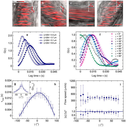
Validation measurements in Zebrafish embryos (3 days post fertilization d.p.f.). (a)–(d) Confocal xy-images acquired by detecting the fluorescence signal (shown in red) of DsRed-expressing RBCs (λexc = 561 nm, detection bandwidth = 575–650 nm), overlaid to (non-confocal) transmitted-light images. fline = 1000 Hz, δx = 0.04 μm, scale bar, 10 μm; γ = 90°, 50°, 0°, −50° in (a), (b), (c) and (d), respectively. γ and v are sketched in the reference Cartesian xy-plane. (e) Exemplifying experimental CCFs for increasing column distance, showing the expected decrease of the peak time for lower (J-I)δx values. (f) Normalized CCFs for γ ∈ [0°, 90°] and (J-I)δx = 6.6 μm, fitted to equation (3); errors are within the size of data points. (g), (h) Experimental CCF peak amplitude (in g) and peak time (in h) for γ ∈ [−90°, 90°] (mean ± standard deviation (s.d.), from n = 7 xy-images), fitted to equation (S.44) and equation (4) (derived in the approximation D = 0 in Supplementary Note 2). Best-fit parameters a = 6.3 ± 0.2 μm in (f) and |v| = 424 ± 11 μm/s in (g). (i) Flow speed |v| recovered from the CCFs fit (open circles, mean ± s.d., n = 4) and |v|0 recovered directly from the CCFs peak time (filled squares, weighted average ± s.d., n = 7). In the lower panel, |v|/|v|0 is shown for γ ∈ [−80°, 80°]. For γ = ± 90°, |v|0 has not been recovered since the CCF turns into a decay (see panel f). |