
Fig. 2 S1
- ID
- ZDB-FIG-171206-22
- Publication
- Kozlovskaja-Gumbrienė et al., 2017 - Proliferation-independent regulation of organ size by Fgf/Notch signaling
- Other Figures
- All Figure Page
- Back to All Figure Page
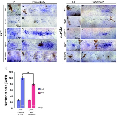
Hippo signaling components in the lateral line. (A–A') In the wildtype lateral line the canonical Hippo pathway inhibitor stk3 (the ortholog of hippo kinase in Drosophila) is expressed in central cells of the primordium, as well as in central cells of deposited neuromasts. (B–B') stk3 is downregulated in NICD and hs:dkk1b (E–E') primordia and neuromasts. (C–C') stk3 is strongly upregulated in mib1ta52b and apcmcr (D and D') neuromasts and primordia. This suggests that the activation status of canonical Hippo pathway is different in NICD and apc mutants. (F) amotl2a, which inhibits Wnt-induced proliferation is not expressed in the deposited neuromast, but in the central region of the wildtype primordium (F'). (G and G') amotl2a expression is downregulated in NICD primordia and in hs:dkk1b primordia where Wnt signaling is disrupted (J and J'). (H and H') amotl2a expression is not affected in the mib1ta52b primordium, whereas it is induced after Wnt upregulation (I and I'), even though proliferation is reduced hs:dkk1b embryos and upregulated in apc mutants. Scale bars are 25 μm. (K) yap1 morpholino injections do not affect primordium size via the activation of p53. Injection of yap1 morpholino into p53 homozygous embryos still leads to a significant reduction in primordium cell number. Error bar represents standard error from four independent experiments (p<0.01=** Student's t test). |