
Fig. 3
- ID
- ZDB-FIG-171206-15
- Publication
- Dzieciolowska et al., 2017 - The larvicide pyriproxyfen blamed during the Zika virus outbreak does not cause microcephaly in zebrafish embryos
- Other Figures
- All Figure Page
- Back to All Figure Page
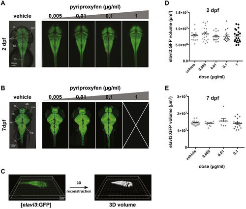
Embryonic and larval brain volumes are not affected by pyriproxyfen treatment. (A) Transgenic [elavl3:GFP] 2 dpf embryo treated with vehicle, 0,005 μg/ml, 0,01 μg/ml, 0,1 μg/ml and 1 μg/ml pyriproxyfen were imaged under a confocal microscope. The morphology of the embryonic brain is not affected by any of the doses. (B) Transgenic [elavl3:GFP] 7 dpf larvae treated with vehicle, 0,005 μg/ml, 0,01 μg/ml, 0,1 μg/ml pyriproxyfen were imaged under a confocal microscope. Of note is that none of the embryos treated with a dose of 1 μg/ml survived past 6 dpf. The morphology of the larval brain is not affected by any of the doses. (C) 3D-volume reconstruction of [elavl3:GFP] embryos using Imaris software (Bitplane) and confocal microscopy. (D,E) Quantification of embryonic (2 dpf) (D) and larval (7 dpf) (E) brain volumes shows no significant differences between vehicle-treated and pyriproxyfen-treated embryos (p > 0.05). (N = 2, n = 7 per condition). sc.: spinal cord; hb.: hindbrain; m/h: midbrain/hindbrain boundary; mb.: midbrain; fb.: forebrain; ot.: optic tectum; tv.: tectum ventricle; ob.: olfactory bulb. |