
Fig. S7
- ID
- ZDB-FIG-171127-17
- Publication
- Sedykh et al., 2016 - Novel roles for the radial spoke head protein 9 in neural and neurosensory cilia
- Other Figures
- All Figure Page
- Back to All Figure Page
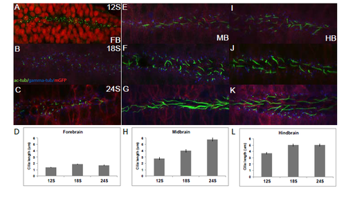
Distinct subpopulations of ventral cilia in each brain subdivision increase in length during neurulation. Tg(β-actin:mGFP) embryos stained with antibodies against acetylated α-tubulin (green), gamma-tubulin (blue) and GFP (red), except in A, where nuclei are stained red. (A-C) Cilia in the ventral FB do not change in length over time. (D) Average length of ventral FB cilia at 12S (1.36μm), 18S (1.87μm) and 24S (1.69μm). (E-G) Ventral MB cilia are longer than primary cilia on average and increase in length over time. (H) Average length at 12S (2.76μm), 18S (4μm) and 24S (5.77μm). (I-K) Ventral HB cilia are longer than MB cilia at 12S and increase in length over time. (L) Average length at 12S (3.72μm), 18S (5.02μm) and 24S (5.01μm). A-C, E-G, I-K are stacked confocal images with anterior to the left. Error bars represent standard error of the mean. |