
Fig. S6
- ID
- ZDB-FIG-171127-16
- Publication
- Sedykh et al., 2016 - Novel roles for the radial spoke head protein 9 in neural and neurosensory cilia
- Other Figures
- All Figure Page
- Back to All Figure Page
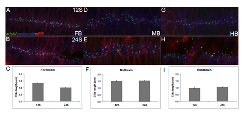
Dorsal cilia are uniform across brain subdivisions and throughout neurulation. Fixed Tg(β-actin:mGFP) embryos stained with antibodies against acetylated α-tubulin (green), gamma-tubulin (blue) and GFP (red). (A,B) Cilia in the dorsal FB do not change in length over time. (C) Average length of dorsal FB cilia at 12S (1.36μm), and 24S (1.02μm). (D,E) Dorsal MB cilia remain unchanged over time. (F) Averaged length at 12S (1.53μm) and 24S (1.54μm). (G,H) Dorsal HB cilia remain unchanged over time. (I) Averaged length at 12S (0.98μm) and 24S (1.07μm). Cilia length was measured in Image J (NIH). Averages were compiled from 20 cilia picked from 10um stacks of the dorsal- and ventral-most regions of the neural tube. A-B, D-E, G-H are stacked confocal images with anterior to the left. Error bars represent standard error of the mean. |