
Fig. S1
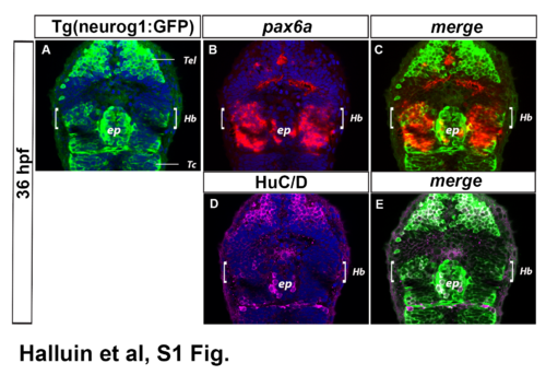
Expression of pax6a and Tg(neurog1:GFP) co-localises in dorsal habenulae. Confocal sections (A-C) of the head of a Tg(-8.4neurog1:GFP)sb1 embryo at 36 hpf after whole-mount immunostaining against GFP (A, green), in situ hybridization against pax6a (B, red) and Immunostaining against HuC/D (D, magenta); cell nuclei staining (in blue) makes brain structures visible in A, B, D, and merges are shown in C (A+B) and E (A+D). The Tg(-8.4neurog1:GFP) transgene is expressed in the epithalamus, both in epiphyseal (ep) and habenular neurons (Hb, white brackets), as well as other brain structures such as the telencephalon (Tel) and the tectum (Tc). The expression of Tg(-8.4neurog1:GFP)sb1 recapitulates endogenous neurog1 expression in habenular progenitors (described previously in [25]), although it can also be detected in newly-born HuC+ habenular neurons, probably due to persistence of the fluorescent reporter which acts as a short term lineage label (E). The expression of pax6a overlaps broadly with most of the Neurog1:GFP+ neurons in both the left and right habenulae (n = 15/15; C). |