
Fig. 3
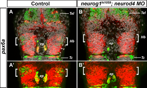
Expression of pax6a is unaffected in neurog1hi1059;neurod4 morphant embryos. Confocal sections (A,B) or 10μm maximum projections (A’,B’) of the head of wild type (A; n = 6/6) or neurog1hi1059;neurod4 Mo injected embryos (B; n = 10/10) at 36 hpf after a whole-mount in situ hybridization against pax6a (red) and an immunostaining against HuC/D protein (green); cell nuclei staining (in grey) makes visible brain structures (A,B). As previously described, the neuronal marker HuC/D is expressed in the epithalamus, both in epiphyseal projection neurons (ep) and in habenular neurons (Hb, white brackets) located on either sides of the epiphysis. The expression of HuC/D is abrogated or strongly reduced in the habenular domain of neurog1hi1059;neurod4 morphant embryos, while it is still detected in the telencephalon (Tel), in the epiphysis and in neurons of the tectum (Tc) (B,B’). On the contrary, no change is seen in the expression of pax6a in the same region, suggesting that the expression of neurog1 and neurod4 does not regulate pax6a during habenular neurogenesis. Embryos are viewed dorsally with anterior up. The expression of HuC/D and pax6a in neurog1hi1059 mutants (n = 5/5) and neurod4 Mo injected embryos (n = 11/11) was similar to that observed in the non-injected controls (data not shown). |
| Gene: | |
|---|---|
| Antibody: | |
| Fish: | |
| Knockdown Reagent: | |
| Anatomical Terms: | |
| Stage: | Prim-25 |
| Fish: | |
|---|---|
| Knockdown Reagent: | |
| Observed In: | |
| Stage: | Prim-25 |