
Fig. 5
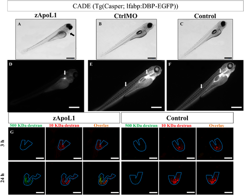
zApoL1 affects the glomerular filtration barrier of zebrafish larvae. After KD of zApoL1, larvae (CET strain) developed edema (arrow in A), a hallmark of a leaky glomerular filtration barrier. The loss of EGFP-vitamin D-binding protein from the blood resulted in a marked decrease of the fluorescence in the blood vessels (D). Only the fluorescence of the podocytes remained (arrow in D). In contrast the control larvae with an intact filtration barrier showed an intense staining of the vessels and the pronephric glomerulus (arrows in E, F). Alexa647-conjugated 10 kDa dextran and FITC-conjugated 500 kDa dextran were injected into the cardinal vene of the larvae (3 dpf). 3 and 24 h after injection, the fluorescence in the pronephric tubules (outlined in blue in G) was determined. Only zApoL1MO larvae showed a green fluorescence in the tubules at 24 h, indicating leakage of 500 kDa dextran through the glomerular filtration barrier. The control larvae showed only the red fluorescence of the filtered 10 kDa dextran. Scale bars represent 500 μm (A-F) and 50 μm (G). |
| Gene: | |
|---|---|
| Fish: | |
| Knockdown Reagent: | |
| Anatomical Terms: | |
| Stage: | Day 4 |
| Fish: | |
|---|---|
| Knockdown Reagent: | |
| Observed In: | |
| Stage: | Day 4 |