FIGURE

Fig. 2

ID
ZDB-FIG-171110-23
Publication
Sun et al., 2016 - Effects of copper oxide nanoparticles on developing zebrafish embryos and larvae
Other Figures
All Figure Page
Back to All Figure Page
Fig. 2

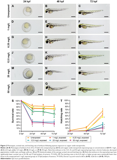
Phenotypes, survival rate, and hatching rate of embryos following aqueous exposure to copper oxide nanoparticles.
Notes: (AR) Phenotypes of embryos from 24 to 72 hpf in the unexposed group (AC) and copper oxide nanoparticle-exposed groups at concentrations of (DF) 1 mg/L, (GI) 6.25 mg/L, (JL) 12.5 mg/L, (MO) 25 mg/L, or (PR) 50 mg/L. Note that embryos in the 12.5, 25, and 50 mg/L exposed groups have a shorter body axis, decreased pigmentation, smaller eyes, and larger yolk sacs. (S and T) Statistical analysis of the average survival rate and hatching rate. (S) Note that embryos in the exposed groups have significantly lower survival rate at 24, 48, and 72 hpf (analysis of variance, *P<0.05). (T) The hatching rates in the 6.25 mg/L or higher groups were statistically lower than those in the unexposed and 1 mg/L exposed groups at 72 hpf (analysis of variance, *P<0.05). Dorsal is up and rostral is left in (AR). Scale bar in (AR): 500 μm.
Abbreviation: hpf, hours postfertilization.

Expression Data

Expression Detail
Antibody Labeling
Phenotype Data

Phenotype Detail
Acknowledgments
This image is the copyrighted work of the attributed author or publisher, and ZFIN has permission only to display this image to its users. Additional permissions should be obtained from the applicable author or publisher of the image. Full text @ Int. J. Nanomedicine