
Fig. S1
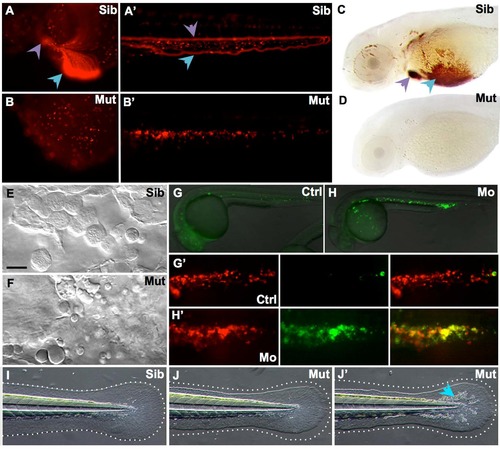
MonNQ039 mutants and Trim33 morphants share phenotypic traits with monTB222 mutants. (A-B') In vivo images of DsRed+ erythroid cells in monNQ039Tg(gata1:DsRed) embryos at 48 hpf in the yolk sac (A,B) or tail (A',B') of sibling (A, A') and mutant (B,B') embryos. Circulating erythrocytes can be seen in the siblings heart (a, purple arrowhead) and duct of Cuvier (A, blue arrowhead), as well as in their caudal artery (A', purple arrowhead) and caudal vein (A', blue arrowhead), but not in the moonshine mutant (B-B'). (C,D) Whole mount O-dianisidine staining of hemoglobin-loaded erythrocytes at 72 hpf in the anterior region of sibling (C) and mutant (D) monNQ039/Tü larvae. Erythrocytes can be seen in the duct of Cuvier (blue arrowhead) and heart (purple arrowhead) of sibling but not mutant larvae. (E-F) In vivo video-enhanced Nomarski microscopy at 18 hpf reveals that the Posterior Blood Island of a monNQ039 mutant (F) is full of apoptotic bodies instead of erythroid progenitors as seen in the sibling (E). (G-H') In vivo images of whole (G,H) or PBI (G',H') of Tg(TBP:Gal4;UAS:AnV-YFP) (G,H) or Tg(TBP:Gal4/UAS:AnV-YFP; gata1a:DsRed) (G',H') embryos at 26 hpf. Embryos injected with the Trim33 morpholino (H,H’) have a lot of apoptotic cells (green channel) corresponding to erythroid progenitors (red channel), unlike in the control embryos (G,G'). (I-J') MonNQ039 mutants (J-J') display a smaller and more irregularly shaped caudal fin than their siblings (I), as shown by the dotted line. Some mutants also have more iridophores (J', blue arrowhead). Ctrl, control; Mo, morphant; Mut, mutant; Sib, sibling. |
| Fish: | |
|---|---|
| Knockdown Reagent: | |
| Observed In: | |
| Stage Range: | 14-19 somites to Protruding-mouth |