
Fig. 3
- ID
- ZDB-FIG-170926-2
- Publication
- Packard et al., 2017 - Automated Segmentation of Light-Sheet Fluorescent Imaging to Characterize Experimental Doxorubicin-Induced Cardiac Injury and Repair
- Other Figures
- All Figure Page
- Back to All Figure Page
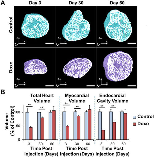
Cardiac Architecture Following Doxorubicin Treatment. Adult zebrafish hearts were harvested at days 3, 30, and 60 following treatment with doxorubicin or control vehicle. (A) Control hearts exhibit preserved architecture throughout the study period. In contrast, doxorubicin-treated hearts demonstrate a profound cardiac remodeling leading to acute decrease in size at day 3, followed by progressive increase at day 30, and return to control levels at day 60. (B) Quantitative analysis of the total heart, myocardial, and endocardial volumes normalized to control values demonstrating the cardiac regeneration process following response to chemotherapeutic injury. **P < 0.01. Doxo: doxorubicin. Scale bar: 200 μm. |