
Fig. 2
- ID
- ZDB-FIG-170926-1
- Publication
- Packard et al., 2017 - Automated Segmentation of Light-Sheet Fluorescent Imaging to Characterize Experimental Doxorubicin-Induced Cardiac Injury and Repair
- Other Figures
- All Figure Page
- Back to All Figure Page
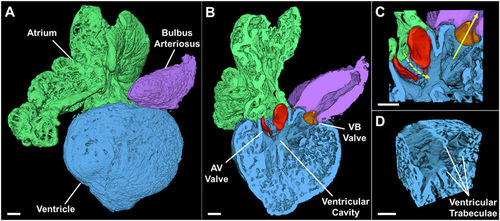
3-D Rendering of the Adult Zebrafish Heart. Example of a reconstructed heart. (A) The anatomic relationship of the intact atrium, ventricle, and bulbus arteriosus is shown in a surface view. (B) A cross-section through the atrium, ventricle, and bulbus arteriosus demonstrates the 2 leaflets of the AV valve (red) and of the VB valve (orange). (C) Magnification at the level of the cardiac valves showing the ventricular inflow through the AV valve (dashed arrow) and the ventricular outflow through the VB valve (solid arrow). (D) Magnification of a ventricular segment with highlighted trabeculae. AV: atrioventricular. VB: ventriculobulbar. Scale bar: 100 μm. |