
Fig. 5
- ID
- ZDB-FIG-170922-52
- Publication
- Xue et al., 2017 - The Vascular Niche Regulates Hematopoietic Stem and Progenitor Cell Lodgment and Expansion via klf6a-ccl25b
- Other Figures
- All Figure Page
- Back to All Figure Page
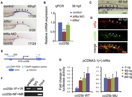
Klf6a Directly Regulates ccl25b Expression (A) Expression of ccl25b in the CHT of control, klf6a morphants, and klf6a−/− embryos at 48 hpf. Black arrowheads mark the ccl25b expressed cells. (B) The qPCR result of ccl25b expression in control, klf6a morphants, and klf6a−/− embryos at 36 hpf (mean ± SD, t test; ∗p < 0.05, ∗∗∗p < 0.001, n = 3). (C) High-magnification imaging of ccl25b expression pattern in the CHT at 48 hpf. Black arrowheads mark the ccl25b expressed cells. The black dashed lines mark the boundary of the caudal artery. Scale bar, 30 μm. (D) Double FISH imaging shows klf6a expression (red), ccl25b expression (green), and co-expression (yellow) in the CHT at 40 hpf. White arrowheads indicate gene expression. Scale bar, 30 μm. (E) The schematic structure of the ccl25b promoter. (F) The ChIP-PCR result shows that Klf6a could directly bind to the promoter of ccl25b. The ccl25b-F/R primers were used to amplify the promoter region containing the conserved Klf6a binding site. The ccl25b-NF/NR were negative control primers used to amplify the ccl25b promoter region without the conserved Klf6a binding site. IgG, immunoglobulin G. (G) Reporter assay shows the regulation of ccl25b promoter by Klf6a. HEK293T cells were co-transfected with the promoter construct of ccl25b containing the conserved Klf6a binding sites (CACCC) or mutated sites (TGTTA) together with the pCDNA3.1(+)-Klf6a plasmid (mean ± SD, t test; ∗p < 0.05, n = 3). See also Figure S5. |
| Genes: | |
|---|---|
| Fish: | |
| Knockdown Reagent: | |
| Anatomical Terms: | |
| Stage Range: | Prim-25 to Long-pec |
| Fish: | |
|---|---|
| Knockdown Reagent: | |
| Observed In: | |
| Stage Range: | Prim-25 to Long-pec |
Reprinted from Developmental Cell, 42(4), Xue, Y., Lv, J., Zhang, C., Wang, L., Ma, D., Liu, F., The Vascular Niche Regulates Hematopoietic Stem and Progenitor Cell Lodgment and Expansion via klf6a-ccl25b, 349-362.e4, Copyright (2017) with permission from Elsevier. Full text @ Dev. Cell