
Fig. 8
- ID
- ZDB-FIG-170829-24
- Publication
- Förster et al., 2017 - An optogenetic toolbox for unbiased discovery of functionally connected cells in neural circuits
- Other Figures
- All Figure Page
- Back to All Figure Page
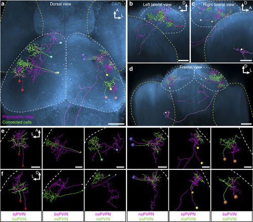
Registration of Optobow fish highlights relative anatomical positions of connected cell pairs. a–d Registration of six identified cell pairs (illustrated by the same cell body color) into one reference brain (outlined by DAPI). Shown are dorsal (a), lateral (b and c) and frontal views (d). Optic tecta are outlined by white, and eyes by yellow dashed lines, respectively. The RGC-PVIN cell pair (white cell bodies) described in Fig. 6 was added in b–d. Scale bar, 50 µm. e, f Dorsal (e) and transverse views (f) of single cell pairs shown in a–d. White dashed lines outline skin. Identified cell types are indicated below. Scale bar, 20 µm |