
Fig. S1
- ID
- ZDB-FIG-170815-48
- Publication
- Förster et al., 2017 - Genetic targeting and anatomical registration of neuronal populations in the zebrafish brain with a new set of BAC transgenic tools
- Other Figures
- All Figure Page
- Back to All Figure Page
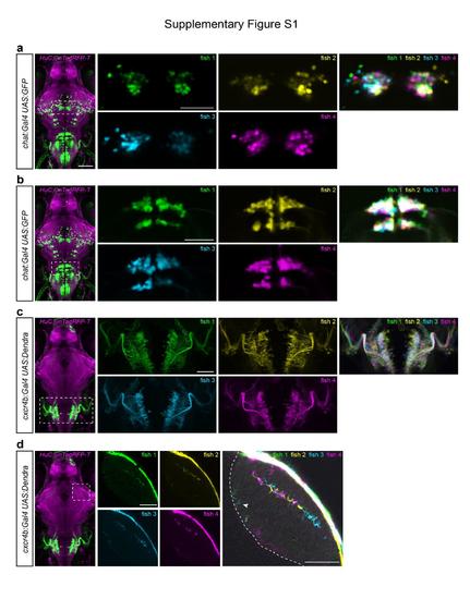
Registration of different larvae of the same transgenic line illustrates variegated, but highly reproducible expression patterns. (a,b) Single confocal slices of chat:Gal4 expression in extraocular motor nuclei (a) and hindbrain neurons (b). After sorting larvae for bright expression of the UAS reporter gene, four different larvae were imaged and co-registered using HuC:lyn-TagRFPT expression as a reference. (c) Z-projection of confocal stacks showing the expression pattern of cxcr4b:Gal4 in the hindbrain. The overlay image reveals that consistent cell body position and axonal projections are largely consistent across four individual fish. (d) Single confocal slices of cxcr4b:Gal4 expression in the tectum. An overexposed overlay is shown on the right. The labeled axons innervate the same layers (SFGS3 and SAC/SPV, arrow head) in the tectal neuropil in all four fish, although their precise location within the layers is variable between the four fish. Dashed line in the right panel indicates the border between the tectal neuropil and cell body layer. Scale bar, 100 μm (whole-brain images on the left) and 50 μm (close-ups). |