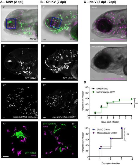
Macrophages are not infected by SINV or CHIKV and not required for neuroinvasion. (A,B) Whole-mount immunohistochemistry of SINV-infected (A) or CHIKV-infected (B) mpeg:G/U:Nfsb-mCherry (macrophages in magenta) larva at 2 dpi. Lateral views of the head; confocal imaging, maximal projection. (A′,B′) Merge of transmitted light with green (infected cells) and magenta (macrophages) fluorescence. (A″,B″) Green fluorescence showing infected cells. (A‴,B‴) Magenta fluorescence showing macrophage distribution. (A‴′,B‴′) Magnification of the area boxed in A′ and B′; green and magenta fluorescence, 3D rendering slightly tilted. Scale bars: 50 µm (A′-A‴,B′-B‴), 25 µm (A″″,B″″). (C) Macrophage depletion in mpeg:G/U:Nfsb-mCherry larvae. Superposition of transmitted light and magenta (macrophage) fluorescence, maximal projection, lateral view of the head, in a DMSO-treated control (top) or 2 days after treatment with metronidazole (bottom). (D) Impact of macrophage depletion on occurrence of neuroinvasion. mpeg:G/U:Nfsb-mCherry larvae were treated with DMSO or metronidazole, before injection with SINV (top graph) or CHIKV (bottom graph). Percentage of brain-infected larvae. n=36 from three independent experiments pooled. ns, not significant (Log-rank test).
|
