
Fig. 5
- ID
- ZDB-FIG-170620-27
- Publication
- Koth et al., 2017 - High-Resolution Magnetic Resonance Imaging of the Regenerating Adult Zebrafish Heart
- Other Figures
- All Figure Page
- Back to All Figure Page
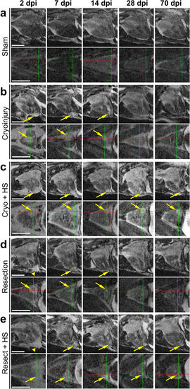
Comparing well and poorly healing hearts for 70 days post injury (dpi). (a–e) Optical sagittal and horizontal MR sections of 5 individual Tg(hsp70l:dnfgfr1a-EGFP) +/− adult fish imaged repeatedly over 70 days showing (a) a sham control ventricle (no injury but opened pericardium), and the regeneration process in (b) cryo-injured and (d) resected ventricles, and (c) cryo-injured plus heat-shock (HS) and (e) resected plus HS treated ventricles at 2, 7, 14, 28, and 70 days post injury (dpi). b + d serve as injured but not HS treated controls to visualise normal versus impaired (c + e) heart repair. Arrows point to the injury location and arrowheads indicate the blood clot following resection injury. Scale bar 1 mm. |
| Fish: | |
|---|---|
| Conditions: | |
| Observed In: | |
| Stage: | Adult |