FIGURE

Fig. 6

ID
ZDB-FIG-170606-24
Publication
Yoo et al., 2017 - Mind Bomb-Binding Partner RanBP9 Plays a Contributory Role in Retinal Development
Other Figures
All Figure Page
Back to All Figure Page
Fig. 6

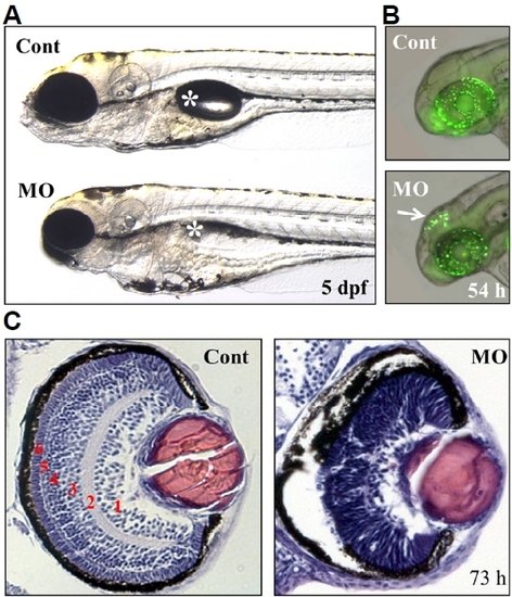
Defects in the brain and retina of ranbp9-MO injected zebrafish embryos.

(A) Control (Cont) and ranbp9-MO injected (MO) zebrafish larvae at 5 dpf. Compared to normal development of body trunk region, reduced size of brain and eye was observed in the MO injected larva. Asterisk indicates swim-bladder. (B) By acridine orange staining, a specific cell death was detected in the brain tectum of 54 hpf morphants. (C) Disruption of retinal development in ranbp9 morphant. Section of HE-stained retina in control and ranbp9 morphant at 73 hpf. In wild-type, the retina was established into several layers; 1, ganglion cell layer: 2, inner plexiform layer: 3, inner nuclear layer: 4, outer plexiform layer: 5, outer nuclear layer: and 6, photoreceptor cell layer, respectively. In morphants, however, these layer formations were not well established.

Expression Data

Expression Detail
Antibody Labeling
Phenotype Data
Fish:
Knockdown Reagent:
Observed In:
Stage Range: Long-pec to Day 5

Phenotype Detail
Acknowledgments
This image is the copyrighted work of the attributed author or publisher, and ZFIN has permission only to display this image to its users. Additional permissions should be obtained from the applicable author or publisher of the image. Full text @ Mol. Cells