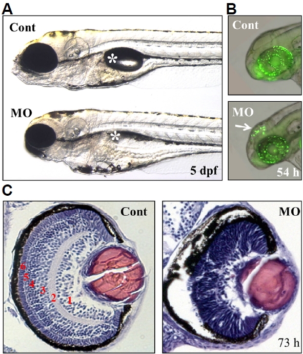
IMAGE

Fig. 6

ID
ZDB-IMAGE-170606-16
Source
Figures for Yoo et al., 2017
Image
Figure Caption

Fig. 6

Defects in the brain and retina of ranbp9-MO injected zebrafish embryos.

(A) Control (Cont) and ranbp9-MO injected (MO) zebrafish larvae at 5 dpf. Compared to normal development of body trunk region, reduced size of brain and eye was observed in the MO injected larva. Asterisk indicates swim-bladder. (B) By acridine orange staining, a specific cell death was detected in the brain tectum of 54 hpf morphants. (C) Disruption of retinal development in ranbp9 morphant. Section of HE-stained retina in control and ranbp9 morphant at 73 hpf. In wild-type, the retina was established into several layers; 1, ganglion cell layer: 2, inner plexiform layer: 3, inner nuclear layer: 4, outer plexiform layer: 5, outer nuclear layer: and 6, photoreceptor cell layer, respectively. In morphants, however, these layer formations were not well established.

Figure Data
Acknowledgments
This image is the copyrighted work of the attributed author or publisher, and ZFIN has permission only to display this image to its users. Additional permissions should be obtained from the applicable author or publisher of the image. Full text @ Mol. Cells