
Fig. 4
- ID
- ZDB-FIG-170601-19
- Publication
- Pietri et al., 2017 - The Emergence of the Spatial Structure of Tectal Spontaneous Activity Is Independent of Visual Inputs
- Other Figures
- All Figure Page
- Back to All Figure Page
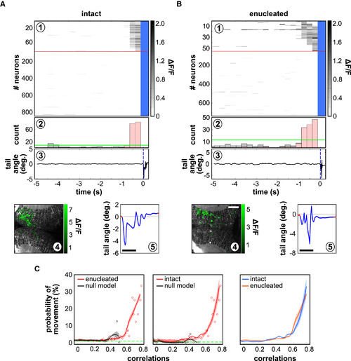
Spontaneous Neuronal Assemblies Are Predictive of Tail Motor Behaviors, even in Chronic Absence of Retinal Inputs (A and B) Examples of spontaneous activation of a topographically compact tectal assembly before the onset of a tail movement in an intact larva (A) and an enucleated larva (B). (A1 and B1) Raster plot of all imaged tectal neurons; the red line separates the neurons belonging to the active neuronal pattern before the tail flip (above), from the rest of the neurons in the circuit (below; notice that the scale is different above and below the line; blue indicates frames during tail-flip movements). (A2 and B2) Histogram of the number of active neurons in the circuit; the fraction of neurons in the active neuronal pattern is represented in pink; the green line represents the threshold for significant neuronal population events. (A3 and B3) Tail angle. The blue dashed line indicates the onset of tail movement. Deg, degree. (A4 and B4) Topography of the spontaneously active neuronal pattern; compactness index: 0.54 (A4) and 0.33 (B4). (A5 and B5) A magnification of the angle of the tail during the movement. Scale bars, 100 ms. (C) Probability of a tail movement as a function of the correlation between all assembly patterns and the spontaneous tectal network activity for the imaging frame preceding the movement onset in intact larvae (left; n = 6) and enucleated larvae (middle; n = 6). Red dots indicate raw data; black dots indicate null models; red curves indicate regression fits, with CI95%; black curves indicate null-model assemblies; and dashed green lines indicate global average probability of movements. Right: comparison between intact larvae (blue) and enucleated larvae (red). |