
FIGURE
Fig. 7
Fig. 7
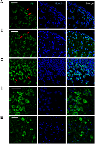
Pin1 subcellular localization in the adult zebrafish brain. Confocal Immunofluorescence analysis on brain coronal sections using Pin1 (green) as primary antibody. Nuclei were stained with Hoechst (blue). Digital magnification of selected areas from images shown in Fig 5 and S6 Fig. (A) lateral zone of dorsal telencephalic area, (B) diffuse nucleus of the inferior lobe, (C) periventricular gray zone of the optic tectum, (D) lateral zone of rostroventral medulla oblongata, (E) central area of caudal medulla oblongata. Scale bar = 25 μm. |
Expression Data
| Gene: | |
|---|---|
| Fish: | |
| Anatomical Terms: | |
| Stage: | Adult |
Expression Detail
Antibody Labeling
Phenotype Data
Phenotype Detail
Acknowledgments
This image is the copyrighted work of the attributed author or publisher, and
ZFIN has permission only to display this image to its users.
Additional permissions should be obtained from the applicable author or publisher of the image.
Full text @ PLoS One