
Fig. S3
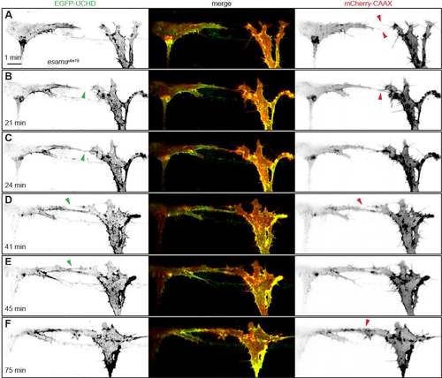
Loss of Esama does not lead to defects in anastomosis (A-F) Still images from Movie S3 of an esamaubs19 mutant Tg(fli1ep:gal4ff)ubs3, Tg(UAS:EGFPUCHD) ubs18, Tg(kdrl:mCherry-CAAX)s916 embryo at around 32hpf, anterior to the left. Single channels are shown in inversed contrast (green is EGFP-UCHD and red is mCherry-CAAX on left and right, respectively) and the merge is shown in the middle. (A) Two tip cells are extending filopodia (red arrowheads) towards each other. (B) A filopodial contact is established (red arrowhead), but actin cytoskeleton (green arrowhead) has not accumulated at the cell-cell bridge yet. (C) The cell-cell bridge is quickly stabilized with actin cytoskeleton (green arrowhead). (D and E) A second protrusion is established between the two tip cells (red arrowhead) and quickly stabilized with actin cytoskeleton (compare green arrowheads in C and D). (F) During contact maturation, the two cell-cell contact sites are fused to a single one (red arrowhead). Scale bar, 10μm. |