
Fig. S2
- ID
- ZDB-FIG-170309-9
- Publication
- Reinig et al., 2017 - The Descending Diencephalic Dopamine System Is Tuned to Sensory Stimuli
- Other Figures
- All Figure Page
- Back to All Figure Page
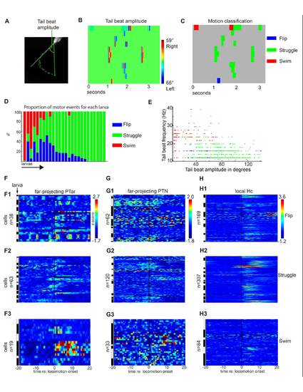
(A-C) Tail motion classification method. (A) The tail beat amplitude of a 4 dpf nacre-/- larva is extracted using the chain of tail midpoints described in Supplemental Figure S1I. (B) Tail beat amplitude heat map in which the x-axis corresponds to time and the y-axis to 11 different portions of the high-speed camera recording. (C) The tail beat amplitude (B) was used for classification of the locomotion patterns flip, struggle and swim. (D) Proportion of the three locomotor patterns in individual larvae. Each bar corresponds to a single recording of one larva. (E) Plotting the tail beat frequency versus tail beat amplitude for all spontaneous motor events recorded during 2-photon calcium imaging experiments. Each motor event is presented as a single dot in blue (flip), green (struggle) or red (swim). Spontaneous slow swim is clearly distinguished from struggle by smaller tail beat amplitude and higher tail beat frequency. (F-H) Median single cell calcium activity as z-score in a 40 second time frame centered around the onset of flip, struggle or swim behavior in the far projecting PTac, PTN and local projecting Hc dopaminergic subgroups. The white and black bars indicate different larvae. |