|
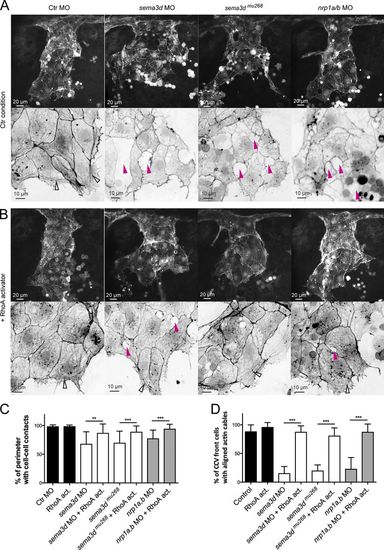
Sema3d-Nrp1 signaling regulates cell–cell contacts and Actin cable formation in the CCV leading edge by activating RhoA. (A and B) Lateral confocal projections of Tg(fli1a:lifeactEGFP)mu240 embryos at 30 hpf. Magnifications of the leading edges were color-inverted. (A) sema3d morphants and mutants as well as nrp1a/b morphants exhibited lesions in the leading edge (pink arrowheads) and lacked parallel-arranged Actin cables (open arrowhead in Ctr MO). (B) Activation of RhoA from 24 to 30 hpf reduced lesions (pink arrowheads) and rescued Actin cable formation (open arrowheads) in sema3d morphants and mutants as well as in nrp1a/b morphants. (C and D) Quantification of cell–cell contact length (C, n = 16) and cells with Actin cables (D, each n = 8; sema3dmu268 + RhoA activator, n = 4). ***, P < 0.001; **, P < 0.01. Error bars indicate SD.
|