
Fig. 7
- ID
- ZDB-FIG-170222-16
- Publication
- Manalo et al., 2016 - Differential Lectin Binding Patterns Identify Distinct Heart Regions in Giant Danio (Devario aequipinnatus) and Zebrafish (Danio rerio) Hearts
- Other Figures
- All Figure Page
- Back to All Figure Page
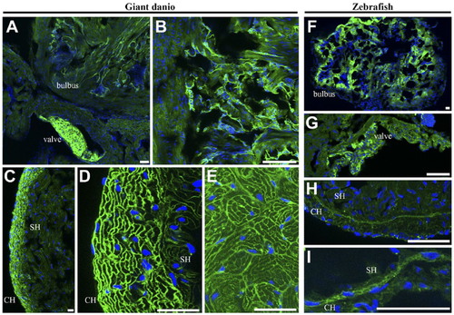
Differential tomato lectin binding in giant danio and zebrafish hearts. Tomato lectin staining was present in the bulbus endothelium and mesenchyme, and strong in the valves (A, B; higher magnification of bulbus) of the giant danio. Binding was also strong in the CH, staining the cardiomyocyte borders (C, D; higher magnification). A similar binding pattern was seen in the SH but weaker (E). In the zebrafish, tomato lectin staining was present in the bulbus (F) and the valve (G). Little to no reactivity was seen in the ventricle except for low but discernable binding in the junctional region (H) and to the borders of the transitional cardiac myocytes (I) bridging the compact and spongy myocardia. Abbreviations: SH, spongy heart; CH, compact heart. Scale bars: A–I = 20 µm. |