
Fig. 2
- ID
- ZDB-FIG-170222-12
- Publication
- Manalo et al., 2016 - Differential Lectin Binding Patterns Identify Distinct Heart Regions in Giant Danio (Devario aequipinnatus) and Zebrafish (Danio rerio) Hearts
- Other Figures
- All Figure Page
- Back to All Figure Page
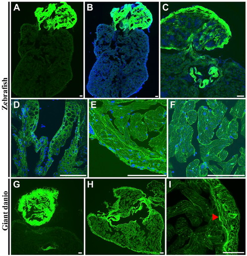
Differential wheat germ agglutinin (WGA) lectin binding in the zebrafish and giant danio hearts. In the zebrafish, the strongest WGA lectin binding was observed in the bulbus mesenchyme (A, B) with the ventricle appearing minimally stained by comparison, and in the valves (C, D). At higher magnification, WGA could be seen staining the compact heart, the trabecular bundles of the spongy heart (E), and the cardiomyocyte borders (F). In the giant danio, the staining pattern was similar with the strongest staining level in the bulbus mesenchyme (G) with the ventricle appearing minimally stained. When imaged without the bulbus, WGA staining is seen throughout the ventricle but with higher levels in the compact heart and the valves (H). At higher magnification, WGA was observed to stain cardiac myocyte borders as well as the junctional region (arrowhead) of the giant danio heart (I). Scale bars: A–I = 50 µm. |