
Fig. S3
- ID
- ZDB-FIG-170220-11
- Publication
- Mayrhofer et al., 2017 - A novel brain tumour model in zebrafish reveals the role of YAP activation in MAPK/PI3K induced malignant growth
- Other Figures
- All Figure Page
- Back to All Figure Page
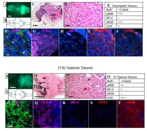
Histological and immunological appearance of IVth ventricle and diencephalic tumours. A) Representative diencephalic tumour in zic:RASsomatic fish. B) Schematic drawing, indicating the position of the sections shown in F-J. C) H&E stained section, boxed area indicates enlargement shown in D. E) Summary of the immunohistochemical observations related to diencephalic tumours. F-J) Immunostaining of heterotopia sections stained as indicated. K) Representative IVth ventricle tumour in zic:RASsomatic fish. L) Schematic drawing, indicating the position of the sections shown in P-T. M) H&E stained section, boxed area indicates enlargement shown in N. O) Summary of the immunohistochemical observations related to IV ventricle tumours. P-T) Immunostaining of IVth ventricle tumour sections stained as indicated. Scale bars: A, K= 2 mm; C, M=200 μm; D, F-J, N, PT = 25 μm. |