
Fig. S5
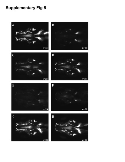
Rescue of atp6v1h mutant zebrafish. Bone staining of uninjected wild type and atp6v1h -/- embryos; loss of bone mineralization is seen in atp6v1h mutant embryos (A and B). atp6v1h mutant embryos at single-cell stage injected with wild type mRNA (C, 200 pg; D, 300 pg) were stained at 5dpf; representative figures demonstrate the rescue of bone phenotype, seen as increase in bone mineralization. atp6v1h mutant embryos injected with mutant mRNA containing the same K386N and N387Y mutations found in humans (E, 200 pg; F, 300 pg) at single-cell stage and stained at 5 dpf; representative figures show the absence of bone staining, indicating that the mutations create a non-functional gene. Wild type and mutant mRNA were injected into wild type embryos at single-cell stage and stained at 5 dpf (G and H); representative figures show no alteration in morphology and bone staining, suggesting the absence of gain-of-function resulting from overexpression of either wild type or mutant mRNA. |