
Fig. S4
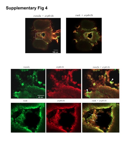
Co-expression analysis of atp6v1h with osteoclast or osteoblast markers. Double fluorescence RNA whole mount in situ hybridization with atp6v1h and rank or runx2a probes were performed on 60 hpf embryos and imaged by confocal microscopy. Upper panels are ventral views of merged lower magnification images of runx2a and atp6v1h or rank and atp6v1h, in which the boxes indicate the regions shown in the lower panels of higher magnification. The lower panels are images of individual colors (green fluorescence for probes of runx2a or rank, red fluorescence for atp6v1h) and merged color (runx2a + atp6v1h, rank+ atp6v1h). Arrowheads point to cells that are mainly positive for runx2a and arrows point to cells that appear positive both for rank and atp6v1h (co-expression: yellow fluorescence). |
| Genes: | |
|---|---|
| Fish: | |
| Anatomical Terms: | |
| Stage: | Pec-fin |