
Fig. 2
- ID
- ZDB-FIG-170125-6
- Publication
- Cepero Malo et al., 2017 - The Zebrafish Anillin-eGFP Reporter Marks Late Dividing Retinal Precursors and Stem Cells Entering Neuronal Lineages
- Other Figures
- All Figure Page
- Back to All Figure Page
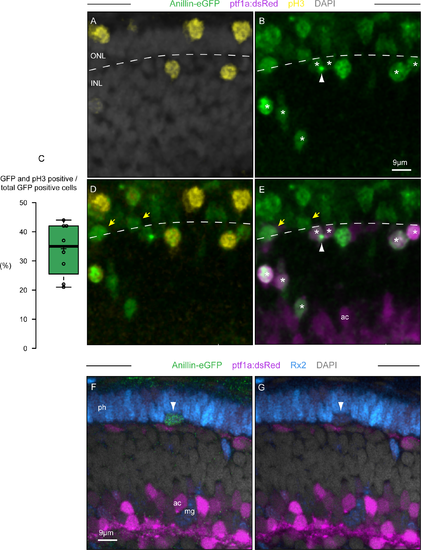
Anillin-eGFP marks all cycling cells of the maturing retinal layers. (A-E) Optical section from a z-stack (frontal view) through the retina of a 48 hpf, anillin:anillin-eGFP/ptf1a:dsRed transgenic zebrafish that have been counterstained with DAPI (A) and pH3-antibody (A,D). (C) Only 34% of the Anillin-eGFP positive cell nuclei are pH3 immunoreactive at 48 hpf (n = 8 retinas). The black horizontal line shows the median while the crosses show the average. The box limits indicate the 25th and 75th percentiles as determined by R software; whiskers extend 1.5 times the interquartile range from the 25th and 75th percentiles, data points are plotted as open circles. Asterisks in (B) and (E) highlight cells that are (ptf1a)dsRed (magenta) and Anillin-eGFP (green) positive, only few of which are also pH3 positive (yellow in D). The arrow in (B) and (E) points at the midbody between dividing daughter cells (asterisk) during late cytokinesis. (F,G) Optical section from a z-stack (frontal view) through the retina of an 60 hpf, anillin:anillin-eGFP/ptf1a:dsRed transgenic zebrafish that have been counterstained with DAPI (grey) and Rx2-antibody (blue). The arrowhead points at the cycling, Anillin-eGFP positive and Rx2-immunoreactive cell located at the base of the outer limiting membrane. ONL, outer nuclear layer; INL, inner nuclear layer; ac, amacrine cell; ph, photoreceptors; mg, Müller glia cell bodies (intermingling with the ac bodies). |