
FIGURE
Fig. 5
- ID
- ZDB-FIG-170104-7
- Publication
- Ryu et al., 2016 - Morphologic Changes of Zebrafish Melanophore after Intense Pulsed Light and Q-Switched Nd:YAG Laser Irradiation
- Other Figures
- All Figure Page
- Back to All Figure Page
Fig. 5
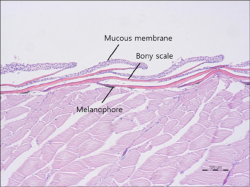
Normal adult zebrafish skin stained with H&E stain at ×100 magnification. Unlike the human skin, zebrafish have scales which are calcified plates originating in the dermis and covered by the mucous membrane. A horizontal view of fish skin reveals melanophores right below the scales. |
Expression Data
Expression Detail
Antibody Labeling
Phenotype Data
Phenotype Detail
Acknowledgments
This image is the copyrighted work of the attributed author or publisher, and
ZFIN has permission only to display this image to its users.
Additional permissions should be obtained from the applicable author or publisher of the image.
Full text @ Ann. Dermatol.