
Fig. 2
- ID
- ZDB-FIG-170104-4
- Publication
- Ryu et al., 2016 - Morphologic Changes of Zebrafish Melanophore after Intense Pulsed Light and Q-Switched Nd:YAG Laser Irradiation
- Other Figures
- All Figure Page
- Back to All Figure Page
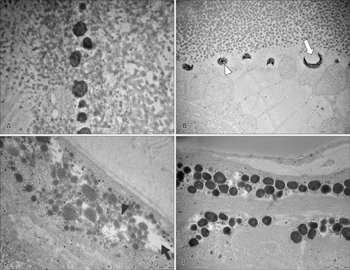
Representative transmission electron microscope images of (A) control, (B) pulse-in-pulse mode intense pulsed light (IPL), (C) IPL irradiation with 560 nm filter and 7 msec, and (D) IPL irradiation with 560 nm filter and 35 msec. (B, C) Irradiation with pulse-in-pulse IPL and conventional IPL for 7 msec induced melanophore thermolysis (black arrowhead), with vacuolization (white arrowhead), central electron lucency (white arrow), and empty spaces (black arrow) evident due to melanophore destruction. Conventional IPL irradiation with 35 msec pulse width caused some melanophore changes but no vacuolization. Scale bar=100 µm. |