
Fig. 3 S1
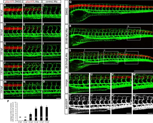
The time course of ectopic blood vessel growth after radial glia ablation, and the emergence of ectopic blood vessels in the trunk regions where radial glia are robustly ablated. (A–O) TgBAC(gfap:gal4ff);Tg(UAS:mcherry-NTR);Tg(kdrl:EGFP) (A and B) and Tg(kdrl:EGFP) (C) trunks at different developmental stages. Animals were treated with DMSO (A) or 2 mM Mtz (B and C) between 30 and 54 hpf and then kept in egg water until confocal analysis. At 60 hpf (A–C), no ectopic vessels were observed in Mtz-treated TgBAC(gfap:gal4ff);Tg(UAS:mcherry-NTR) animals. Note that while TgBAC(gfap:gal4ff);Tg(UAS:mCherry-NTR) expression is significantly decreased in Mtz-treated TgBAC(gfap:gal4ff);Tg(UAS:mcherry-NTR) animals at this stage, some expression remains visible. At 80 hpf (D–F), when TgBAC(gfap:gal4ff);Tg(UAS:mCherry-NTR) expression is dramatically decreased in Mtz-treated TgBAC(gfap:gal4ff);Tg(UAS:mcherry-NTR) fish, ectopic sprouts between ISVs begin to appear. At 100 hpf (G–I) and 130 hpf (J–L), more and more ectopic sprouts have emerged between ISVs and fused with one another or with other ISVs. At 154 hpf (M–O) and later, a majority of the ectopic vessels have apparently fused with one another or with other ISVs and get stabilized in the dorsal part of the trunk. Scale bar, 100 µm. (P) Quantification of average number of somites that showed ectopic blood vessels at different stages of development after radial glia ablation (10 somites examined per animal; 20 animals examined per condition). Animals were treated with 2 mM Mtz between 30 and 54 hpf and then subject to confocal analysis. Values represent means ± SEM. (Q–S) 154 hpf TgBAC(gfap:gal4ff);Tg(UAS:mcherry-NTR);Tg(kdrl:EGFP) trunks treated with DMSO (Q), 2 mM Mtz between 30 and 80 hpf (R), or 2 mM Mtz between 30 and 54 hpf (S). Scale bar, 500 µm. (T–V and T’–V’) High magnification images of selected areas (outlined in white) of the trunk shown in S. Ectopic vessels emerged in the anterior (T and T’) and medial (U and U’) part of the trunk, where radial glia ablation was more efficient. However, ectopic vessels were not observed in the posterior part of the trunk (V and V’), where radial glia ablation was less efficient. Scale bar, 100 µm. (W and W’) High magnification images of a selected area (outlined in white) of the trunk shown in R. Under this ablation treatment condition, the posterior part of the trunk showed more efficient ablation of radial glia than in the one shown in panels (V and V’) and exhibited ectopic vessel sprouting. Scale bar, 100 µm. |