
Fig. 2
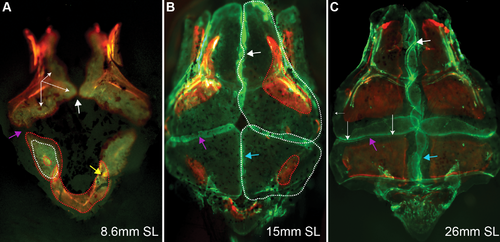
The growth pattern of calvaria bones as revealed by sequential staining with Alizarin red and Calcein green. (A) Initial directions of frontal bone growth (white arrows) and radial growth of the parietal bone are depicted after each vital staining by red and white dotted lines. The supraoccipital bone is depicted by the red dotted line. (B-C) The white, blue and purple arrows indicate developing sutures: interfrontal, sagittal and coronal, respectively. (B) The contour of frontal and parietal bones growth is outlined by red dotted lines labeling the first vital staining with Alizarin and by white-dotted lines the second treatment by Calcein green. (C) Posterior frontal bone advancement (long white arrows) revealed after second vital staining with Calcein green, and lateral growth of frontal bone (small arrow on the left side). Note the interdigitation of the frontal and parietal bones. Skulls were dissected and mounted for imaging from a dorsal view. |