|
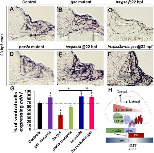
Gsc and Pax2a differentially regulate cdh1. (A–F) Cross-sections passing through the neurogenic domain of the otic vesicle just posterior to the utricular macula showing cdh1 expression at 23 hpf in embryos with indicated genotypes. The otic epithelium is outlined in each image. Arrows in A indicate cells with very low cdh1 expression interspersed with cells showing high cdh1 expression. (Magnification: A–F, 640×.) (G) Means and SD of the percentage of cells in the otic floor expressing cdh1 in the embryos with indicated genotypes. Data were obtained by counting the number of stained and unstained cells in serial sections (n = 3–4). Transgenic embryos were heat shocked at 22 hpf. Asterisks indicate significant differences between groups indicated by brackets or compared with control embryos (P < 0.05). (H) A model for regulation of epithelial tissue dynamics during otic neurogenesis. See Discussion for details.
|