|
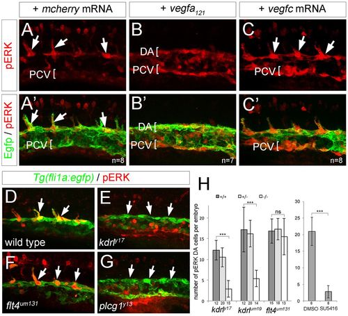
ERK phosphorylation in sprouting ISVs requires Vegfa signaling. (A-G) Confocal images of Tg(fli1a:egfp)y1 embryos at 21 hpf (A-C′) and 20 hpf (D-G) immunostained for GFP (green) and pERK (red). Lateral views, anterior to the left, dorsal is up. Number of analyzed embryos is indicated, representative images are shown. Embryos were injected with 100 pg mcherry mRNA (A,A′), 25 pg vegf121 mRNA (B,B′) or 100 pg vegfc mRNA (C,C′). (D) Wild-type embryo. (E) kdrly17 mutant embryo. (F) flt4um131 mutant embryo. (G) plcg1y13 mutant embryo. Arrows indicate pERK-positive cells, or lack thereof. (H) Graph showing number of pERK+ endothelial cells per embryo at 20 hpf in a defined segment of the DA in kdrl and flt4 mutant embryos, or in embryos treated with 10 µM SU5416. Number of embryos for each genotype or treated with compound is indicated on the x-axis. Error bars represent s.d.; ***P<0.001; ns, not statistically significant.
|